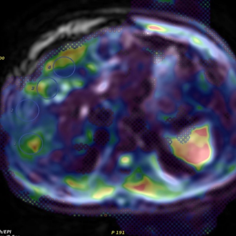
Magnetic Resonance Imaging and Spectroscopy
We develop computer graphics software for creation of 3D, rotating, shaded surface displays of organs and lesions detected in the usual tomographic MR images, special radio frequency pulse sequences and data manipulation software for enhancement of contrast in tomographic images, development of fast MR imaging methods that utilize prior knowledge of anatomy, development of new methods of acquiring and analyzing functional MR images, use of MR to measure tumor metabolism, blood flow, and response to therapy. We also develop an in vivo current density MRI technology to measure the distribution of the current density within the body when an external current source is applied.
Functional Magnetic Resonance Imaging (fMRI)
The development of functional MRI (fMRI) technology is aims for the understating of the neuronal activity of the human brain in the control of the body movements and in performing various cognitive tasks. The experimental platform for our fMRI research program is based on a high filed and research-dedicated MRI scanner. We develop and optimize both fMRI data acquisition and data analysis strategies to better localize the neuronal activity accurately. Cerebral blood flow, blood volume, and tissue oxygenation during brain stimulation has been measured and evaluated. Diffusion tensor imaging is being developed as one of biomarkers for determination of brain disorder that associated with white matter damage.
Research Faculty

Professor of Radiology, Radiation and Cellular Oncology

Maryellen Giger, PhD
A.N. Pritzker Professor of Radiology

Kunoi Doi, PhD
Professor Emeritus