Imaging Modality Chart

Research Overview

The Department of Radiology has 14 basic science faculty members with imaging science research interests including evaluation methodologies & ROC analysis, computer-aided diagnosis & machine learning/deep learning analysis for the interpretation of a variety of medical images (such as breast, thoracic, colon, cardiac, skeletal & radioisotope images), new acquisition methods for MRI and MRIS, novel tomographic reconstruction methods, new methods for PET, SPECT and optical imaging, and developments of novel imaging instrumentation. The faculty labs include over seventy grant-supported researchers including research professors, research associate professors, research assistant professors, research lab computer scientists & staff, research associates, post-docs, graduate students, medical students, undergraduates. A broad range of research activity is under way in the department. We are proud to be among the top twelve Academic Radiology Departments based on NIH funding.

Quick Links

  • 14

    Basic Science Faculty

  • 70+

    Grant-Supported Researchers

  • 3

    Floors in our NIH Funded Research Center

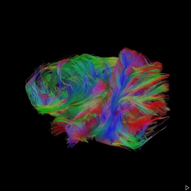
The world's first use of Tc-99m for medical imaging was demonstrated at the UC. Theoretical and application developments for physical image quality assessment of spatial resolution, noise, and contrast were investigated for both analog and digital imaging systems. Research & developments in medical decision making & ROC analysis have benefited researchers around the world through the free availability of state-of-the-art ROC analysis software. Image co-registration and integration research done at UC in the 1980s germinated the field of multi-modality imaging. University of Chicago pioneered the field of computer-aided diagnosis, developing the first prototype for mammographic CAD in the early 1990's. Ground-breaking developments in tomographic image reconstruction have been made yielding analytic solutions to the complex 3D and 4D problems. More recently, developments in small-animal MRI have led to the first image of mammary DCIS in a mouse model. In addition, research at the UC includes solving important problems in muscle and cardiovascular physiology such as NMR detection of gene expression.

The Radiology Research Imaging Center, which occupies three floors and was partially financed by an NIH construction grant, provides the department with a state of the art multidisciplinary radiology imaging research center designed to facilitate greater integrated research and to encourage collaboration across department lines.

In coordination with the Human Imaging Research Office, "de-identified" medical images are provided to researchers spanning multiple disciplines to further scientific endeavors. Additionally, the Brain Research Imaging Center, a joint enterprise between Radiology and Neurology has a 3T and 1.5T MRI equipment to support this endeavor.  The Department is also proud to run the Molecular Imaging and Cyclotron Facility with multiple interdisciplinary projects.  Most recently, the faculty received an NIH Shared Instrument Grant to support high performance computing, NIH S10 OD025081 (Giger), namely the Protected Radiomics Analysis Commons for Deep Learning in Biomedical Discovery, which is currently installed in a machine learning facility within the Department of Radiology, with a reach of 1.9 PFLOPS.

Research Programs and Cores

Computer-aided diagnosis (CAD)/Machine Learning/AI is a broad concept that integrates image processing, machine learning/deep learning, computer vision, mathematics, physics, and statistics into computerized techniques that assist radiologists in their medical decision-making processes.

Read More 

The Department of Radiology has a rich history in developing quantitative methods for evaluating and optimizing radiographic systems. Faculty members in the Department helped to introduce the concepts of modulation transfer function, Wiener (noise power) spectra, and detective quantum efficiency in medical imaging, developed standard methods for measuring these metrics, and demonstrated their utility over a number of different applications such as mammography, skeletal and thoracic radiography. These concepts have been instrumental in the development and optimization of x-ray imaging technologies, including digital radiography.

Read More 

The development of functional MRI (fMRI) technology is aims for the understating of the neuronal activity of the human brain in the control of the body movements and in performing various cognitive tasks. The experimental platform for our fMRI research program is based on a high filed and research-dedicated MRI scanner. 

Read More

The X-ray tomographic imaging research effort at the University of Chicago is an integrated program of system design, hardware optimization, and image reconstruction advances.

Read More

Functional and molecular imaging at the University of Chicago includes a broad spectrum of research agenda in positron emission tomography (PET), singlephoton emission computed tomography (SPECT), x-ray computed tomography (CT), ultrasound, optical imaging (fluorescence and bioluminescence imaging), photoacoustic imaging, etc., in order to enable and improve quantitative measurements of physiologic functions and assessments of molecular pathways, both in health and in disease. 

Read More

Several Radiology faculty are working to develop new methodology for the evaluation of diagnostic performance. Much of this research focuses on Receiver Operating Characteristic (ROC) analysis, which describes the accuracy of each diagnostic modality in terms of the trade-offs that are available between its sensitivity and specificity in a particular clinical task [1-7]. 

Read More

The Human Imaging Research Office is part of the Imaging Research Initiative (IRI) at the University of Chicago. Its goal is to act as the facilitator for investigators with research projects and clinical trials that require imaging-related services and data. 

Read More

Integrated Small Animal Imaging Research Resource (iSAIRR) is a synergistically integrated infrastructure that offers a broad spectrum of imaging modalities, techniques, and services for in vivo imaging of small animals and ex vivo imaging of tissue/organ specimens. Our goal is to provide University of Chicago investigators and external users with state of the art, quantitative multi-modality imaging technologies to advance in-vivo molecular and physiological research of a broad range of disease and cancer models, and to accelerate pre-clinical development of novel therapeutics and diagnostic tools. Currently,

Read More

Development of methodology for evaluation of diagnostic performance has been an essential part of our department’s research for many decades. The primary focus of this work has been on Receiver Operating Characteristic (ROC) analysis, which describes the accuracy of a diagnostic modality, for a particular clinical task, in terms of its trade-off between sensitivity and specificity [2-4]. 

Read More

Research Resources